Category Archives: Service

Laparoscopic Fundoplication

Laparoscopic Fundoplication in Delhi
Laparoscopic fundoplication is a keyhole procedure performed for patients with severe acid reflux, Barrett’s oesophagus and symptomatic hiatus hernias that no longer respond to medication. It is done to prevent acid from the stomach travelling the wrong way into the oesophagus. The operation is predominantly laparoscopic although very occasionally, conversion to open surgery may be necessary.

The procedure involves narrowing the defect through which the oesophagus passes into the stomach (called the hiatus) as well as wrapping the upper part of the stomach around the lower oesophagus (the wrap) to recreate the valve at the lower end of the oesophagus. The wrap itself may be complete or partial depending on the circumstances. The operation takes about 1.5 to 2 hours and is carried out under general anaesthesia.

Overnight hospital stay is to be expected, although some patients can go home on the same day. There are restrictions to food intake for the first few weeks after the operation. Sloppy and pureed food is advised for 6 weeks following the operation. Bread and meat are likely to cause the most trouble and are to be avoided. Dietary advice will be provided before you go home. Anti acid medication can be stopped immediately after the procedure

Hiatal Hernia

hiatal-hernia-treatment-in-delhi
A hiatal hernia occurs when a part of the stomach protrudes through the diaphragm and into the chest. The diaphragm is a sheet of muscle that separates the abdomen from the chest.

The hernia protrudes through the diaphragm at a place called the hiatus. This is the opening in the diaphragm through which the esophagus passes. The esophagus is the tube that connects the throat to the stomach.

There are three main types of hiatus hernias: sliding, para esophageal and mixed. Most people who have a hiatal hernia have a sliding hiatal hernia.

What causes a hiatus hernia?

A hiatus hernia is usually caused by weak muscles and tissue in and around the hiatus.

In a sliding hiatus hernia, a small portion of the stomach pushes up through the diaphragm into the chest. A valve between the esophagus and the stomach also moves upwards away from the diaphragm.

What are the symptoms?

Most people with a hiatus hernia have no symptoms.

One symptom you may have is heartburn, which is an uncomfortable burning sensation, heat or pain behind the breast bone. It is common to have heartburn at night when you are trying to sleep.

If you have symptoms frequently or if they are severe, you may have gastroesophageal flux disease(GERD). A hiatus hernia can cause GERD, and people often have both conditions at the same time.

If you have pain behind your breast bone, it is important to make sure it is not caused by a heart problem. The burning sensation caused by GERD usually appears after eating. Heart pain usually feels like pressure, heaviness, weight, oppression, constriction, discomfort or dull pain. It usually occurs after you have been active.

How is hiatal hernia diagnosed?

A hiatus hernia is usually diagnosed at a doctor’s office or when you are tested for another health problem.

If you have symptoms, your laparoscopic surgeon in Delhi will ask about these. If you have severe or frequent symptoms, you may have gastroesophageal reflux disease(GERD). In this case, your doctor may do more tests or give you medications to treat GERD.

How Is Hiatal Hernia Treatment in Delhi Done?

If you have no symptoms, you do not need hernia treatment in Delhi.

If you have mild symptoms, your doctor may suggest making changes in your lifestyle and possibly taking over-the-counter medications. Here are some things to try:

  • Change your eating habits.
  • After eating, wait for 2 to 3 hours before bedtime. It is not a good idea to eat snacks late at night.
  • Chocolate, mint, and alcohol can make GERD worse. The serelax the valve between the esophagus and the stomach.
  • In some people, very spicy foods, foods that have a lot of acid(such as tomato sand oranges) and coffee can make GERD symptoms worse. If your symptoms get worse after eating certain food, you may want to stop eating that food to see if your symptoms improve.
  • If you have heartburn during the night, raise the head of the bed from 6inches(15cm) to 8inches(20cm) by placing blocks under the bed frame or a foam bed under the head of the mattress. (Using additional pillows does not work).
  • Lose weight, if you need to. Losing just between 5 and 10 pounds(2.3to4.5kg) can help.

If you have frequent symptoms or if the symptoms are severe, you may have GERD. Lifestyle changes may help, and your surgeon in Vikaspuri may prescribe medications. In severe cases, surgery can be done to place the hernia back in the abdomen.

Gallbladder Stone

gallbladder-stone-surgery-in-Delhi
The laparoscopic gallbladder stone surgery in Delhi removes the gallbladder and gallstones through several cuts (incisions) in the abdomen. The laparoscopic surgeon in Delhi will inflate your abdomen with air or carbon dioxide to see clearly.

The surgeon will insert an illuminated instrument attached to a video camera (laparoscope) through an incision near the navel. Next, the surgeon will use a video screen as a guide while inserting surgical instruments into the other incisions to remove the gallbladder.

Gallbladder-stone-treatment-in-Delhi

Before the surgeon removes the gallbladder, you may be given a special x-ray procedure called intraoperative cholangiography, which shows the anatomy of the bile ducts.

You will need general anesthesia for this surgery, which usually lasts 2 hours or less.

After the gallbladder stone surgery in Delhi, bile flows from the liver (where it occurs) through the common bile duct and into the small intestine. Because the gallbladder has been removed, the body can no longer store bile between meals. Most people have little or no digestive effect.

In 5 to 10 of every 100 laparoscopic gallbladder surgeries in India, the surgeon needs to switch to an open surgery method that requires a larger incision. Examples of problems that may require open surgery instead of laparoscopic surgery include unexpected swelling, scar tissue, injury or bleeding.

What to expect after surgery?

You may have gallbladder stone treatment in Delhi on an outpatient basis or you may stay in the hospital for 1 or 2 days.

Most people can return to their usual activities between 7 and 10 days later. Those who undergo laparoscopic gallbladder surgery are sore for about a week. But after 2 or 3 weeks they have much less discomfort than those who had open surgery. No special diets or other precautions are necessary after this surgery.

Why is it done?

Laparoscopic gallbladder stone treatment in Delhi is the best method of treating gallstones that cause symptoms unless there is a reason why this surgery should not be performed.

Laparoscopic surgery is used more frequently when there are no factors present that could complicate it.

Effectiveness

Laparoscopic gallbladder stone surgery in Delhi is safe and effective. The surgery removes the gallstones that are found in the gallbladder. It does not remove the stones present in the common bile duct. Stones can form in the common bile duct years after the gallbladder has been removed, although this is rare.

Risks

The overall risk of laparoscopic gallbladder surgery is very low. The most serious complications that may occur include:

  • Infection of an incision.
  • Internal bleeding
  • Injury to the common bile duct.
  • Lesion to the small intestine caused by one of the instruments used during surgery.
  • Risks of general anesthesia.

Other uncommon complications may include:

  • Gallstones that remain in the abdominal cavity.
  • Bile filtering inside the abdominal cavity.
  • Injury to the blood vessels of the abdomen, such as the main blood vessel that carries blood from the heart to the liver (hepatic artery). This is rare.
  • A gallstone that is pushed into the common bile duct.
  • A cut in the liver.

It may take more than one operation to repair these complications.

After gallbladder removal surgery in Delhi, some people have persistent abdominal symptoms, such as pain, swelling, gas, and diarrhea ( postcholecystectomy syndrome).

To think

Recovery is much quicker and less painful after laparoscopic surgery than after open surgery.

  • The hospital stay after laparoscopic surgery is shorter than the stay after open surgery. People usually return home the same day or the next day compared to the stay of 2 to 4 days or longer for open surgery.
  • Recovery is faster after laparoscopic surgery.
  • After laparoscopic surgery, you will spend less time away from work and other activities (around 7 to 10 days compared to 4 to 6 weeks).
obstructive-jaundice-treatment-in-delhi

Obstructive Jaundice

obstructive-jaundice-treatment-in-delhiAccumulation of bile pigments in the tissues, which in the examination can be seen by the yellow tone of the skin and the conjunctiva, which occurs when there is an obstacle, to the evacuation of the bile, in the bile canaliculus under the microscope (for example, due to primary biliary cirrhosis), in the intrahepatic bile ducts (for example, by sclerosing cholangitis) or in the main bile ducts (for example, choledocholithiasis, tumors, etc.).

The latter group of pathologies are the most frequent and are diagnosed by an increase in bilirubin in the blood, with a predominance of the conjugate or direct form, and the dilation of the bile duct on ultrasound.

Obstructive Jaundice Treatment in Delhi

  • Acute cholangitis: Resuscitation, antibiotics and bile duct drainage.
  • Cholecystectomized with residual choledocholithiasis: Ideally endoscopically.
  • Malignant obstructions: Cancers of the head of the pancreas or of the ampulla of Vater or of the distal bile duct ideally in the surgical form if curative surgery can be offered. In very advanced tumors, endoscopic or percutaneous prostheses are preferred. Patients in intermediate conditions, surgical palliative treatments.

Inguinal Hernia

Inguinal-Hernia-Treatment-in-Delhi
A hernia occurs when an organ or part of it leaves the cavity that contains it and passes through a hole or weak area in one of the layers of the abdominal wall that surrounds the muscle, called the fascia. In the case of inguinal hernias, they are bumps in the groin. This type is more common in men and can develop to lower the scrotum. Sometimes it becomes very painful so inguinal hernia treatment in Delhi is urgently required.

About 75 percent of all hernias occur in the inguinal region.

Causes

Normally, the factors that lead to the development of inguinal hernias are defects of congenital origin or acquired defects.

In the first case, they are caused by a lack of closure of the canal through which it descends from the abdominal cavity to the scrotum (or the round ligament in the female sex), which makes it possible a hernia of the bowel handles.

On the other hand, the realization of efforts when defecating, coughing, lifting heavy objects, etc., can lead to trauma or weakening of the inguinal wall and, therefore, to the formation of hernias.

Symptom

In general terms, the patient suffering from an inguinal hernia has a lump or swelling in this region, although symptoms usually do not occur.

In some cases, mild pain or discomfort is also associated, which is accentuated by efforts. When the enlargement of the hernia occurs, it can get stuck inside the hole and lose blood flow. At this time, it is common for the patient to have a picture of nausea and vomiting and not be able to eliminate gas or have bowel movements. If this happens, immediate hernia surgery in Delhi is required.

Prevention

To prevent the occurrence of hernias, both inguinal and of any kind, it is recommended that the patient use appropriate techniques to lift the weight, avoid or relieve constipation by eating fiber-rich foods and drinking plenty of fluids.

Types

There are two types of inguinal hernias:
Direct inguinal hernia: This type is the one that occurs in the final area of ​​the muscle, next to the opening of the inguinal canal. They usually occur in men over 40 and usually occur due to aging or injury.

Indirect inguinal hernia: It is the most common type in men and occurs when an intestinal loop presses against the inguinal canal. This area may have a congenital defect, which implies a risk factor for hernias to appear over time. Both men and women can suffer from it.

Diagnosis

When diagnosing an inguinal hernia, the laparoscopic surgeon in Delhi can see or feel it through physical examination. You can also request that the patient cough, bend over or lift something. It may not be easily identifiable in babies or children.

In the event that it is not visible, an ultrasound will be requested to determine the diagnosis and location of the hernia, if any. If there is an obstruction of the intestine, an X-ray of the abdomen will probably be performed.

Inguinal Hernia Treatment in Delhi

The surgery is the only hernia treatment in Delhi that will provide the patient with a permanent repair, but maybe a risk factor in people with serious health problems. This treatment repairs weakened tissue and close any holes, a procedure that can sometimes be done with patches of synthetic tissues (meshes).

Other data

The people who are considered more likely to develop a hernia are those who have a family history where they have occurred, with cystic fibrosis or cryptorchidism and, in addition, those who suffer from overweight or enlarged prostate. Hernias may occur at birth, but the bump may not be noticeable until a few years have passed, especially in the case of inguinal hernias, which is more common in children. Even so, they usually do not show symptoms until they are adults.

Ventral Hernia

ventral hernia treatment in Delhi
A ventral hernia or incisional hernia is the result of poor healing of an incision (cut) made in the abdominal wall during surgery. The result is a defect (hole) in it, where abdominal contents (intestinal handles) can come out causing obstructions, pain and, in the most severe cases, intestinal necrosis.

Causes of ventral hernia

There are several causes of ventral hernia and may depend on the patient, the surgery itself, or the surgeon.

Causes related to the patient:

With respect to the patient, there are circumstances that may hinder the correct healing of the surgical wound: obesity, poor nutrition, deficits of some vitamin, collagen diseases (the protein responsible for “manufacturing” the scar), corticosteroid and immunosuppressive treatments.

Causes related to surgery:

With respect to the surgery itself, complications can occur that affect the adequate closure and subsequent wound healing: infection of the wound due to contaminated surgeries, wall defect and closure with excessive tension on the edges.

Causes related to the surgeon:

The most important factor related to good healing of a surgical wound is the proper closure of that incision by the surgeon. What is called a GOOD TECHNICAL closure of the abdominal wall? There is a scientific medical consensus on how this ideal closure technique should be carried out to minimize the subsequent development of a ventral hernia.

The ventral hernia, a serious and common problem The ventral hernia is a very serious problem. In several published articles it is estimated that up to one-third of the surgical interventions in which an incision is made in the abdominal wall present later events.

Even laparoscopic surgery does not get rid of this problem since, although much smaller incisions are made, the frequency of ventral hernia is around 15%.

Ventral Hernia Treatment in Delhi

The ventral hernia treatment in Delhi is surgical. We know that the more surgeries performed, the worse the results. Hence the importance of performing adequate and lasting surgery, performed by an expert surgeon and in a referral center.

The popularization of the use of surgical meshes (prostheses) to treat this problem in a more lasting or permanent way has meant a very important advance. The majority of the scientific community considers its use necessary because its advantages (much better results) far outweigh its disadvantages (discomfort from a foreign body, infection).
The other very important factor that has improved the results has been the dedication of a group of surgeons to develop new, varied, and better surgical techniques in the treatment of ventral hernia in the abdominal wall, especially in the last 15-20 years. To this, laparoscopic surgery has been added as another very useful technique in certain groups of patients.

The importance of a good surgeon to treat ventral hernia For these reasons; its high incidence, its technical difficulty, and the need for an accurate and updated knowledge of anatomy and new surgical techniques, it is fundamental to choose a surgical dedicated to the abdominal wall for the treatment of primary ventral hernia or that has been operated on several occasions ( relapsed). It is crucial to get the best results.

Umbilical Hernia

umbilical hernia treatment in Delhi
A hernia consists of the exit to the outside of an organ covered with peritoneum (membrane that covers the organs of the abdomen), which is usually part of the small intestine or the layers that surround it, through a defect or a hole that is Produces in the abdominal wall, just below the skin. This exit is manifested in the patient’s body as a kind of lump that is palpable and is perceived visually.

Abdominal hernias can be of different types: inguinal, umbilical, femoral or epigastric, among others, depending on what area of ​​the body they are in. In this case, umbilical hernias are those that appear, as the name implies, in the navel or around it.

Types of umbilical hernias

Umbilical hernias, depending on their origin or etiology, can be of two types:

  • Primary umbilical hernia: these types of hernias are those that are present in patients from the moment of their birth and the main reason for their appearance is a defect in the abdominal wall and in the umbilical cord since their formation in the maternal uterus began.
  • Incisional umbilical hernia: these umbilical hernias arise as a cause of previous surgical interventions in the area and that, over the years, the suture or internal surgical wound has been weakened. With continuous efforts in the abdominal area, the hernia finds a place to protrude or go outside.

Causes of the appearance of an umbilical hernia Apart from the causes already mentioned in the previous section, incisional umbilical hernias can have their cause in:

  • Performing strenuous physical exercise.
  • Lifting of very heavy objects.
  • Having chronic constipation and, therefore, making efforts to defecate.
  • Having a chronic cough
  • Enlarged prostate, effort to urinate.
  • Being overweight.

In general, all kinds of activities or health problems that increase pressure in the abdominal area may be susceptible to causing an umbilical hernia. On other occasions, hernias are present from birth, only they do not manifest until more advanced ages.

Umbilical hernia and pregnancy

During pregnancy, the most common hernia that occurs in women is the umbilical hernia. Contrary to what one might think, umbilical hernia in pregnant women does NOT pose any risk or complication for pregnancy or for the fetus.

In pregnant women, umbilical hernias usually occur during the second week of pregnancy due to the increase in the size of the uterus, although it can also occur after giving birth, due to the great effort made. Normally, hernias are usually prior to pregnancy due to, as we mentioned before, a defect in the abdominal wall that in this case becomes evident due to the pressure exerted on the abdomen during pregnancy. As for the surgical treatment of hernias in pregnant women, they should always be operated once they have given birth, since complications could arise during pregnancy.

Symptoms of an umbilical hernia

Usually, umbilical hernias have no symptoms beyond the bulge or belly button. This lump is soft to the touch and, if we exert pressure on it, it is reintroduced inside the abdomen, although it will return to bulge when we stop pressing. In some cases, the patient may feel pain or burning in the area of ​​the hernia when making efforts in the abdomen, such as coughing, sneezing, lifting heavy objects, etc. When this pain manifests, the specialist usually recommends an abdominal girdle, especially for pregnant women.

In general, when the patient is at rest, the umbilical hernias do not cause discomfort. Those types of hernias that are too bulky for the hernia exit orifice may remain what is called “strangled” by the exit orifice. When this occurs, it can prevent proper blood circulation and even ischemia and necrosis in the tissue. Strangulated hernias should be communicated immediately to the specialist so that he can operate them urgently since they are very dangerous for the proper functioning of the organism.

Diagnosis of an umbilical hernia

When the umbilical hernia reaches a considerable size, its diagnosis by the specialist is easy, since it is easily recognizable by physical examination. It is also common to perform diagnostic tests such as CT or ultrasound for the diagnosis of smaller abdominal hernias, since larger abdominal hernias can be seen with the naked eye, although it is not usually done if there are no discomforts in the area.

Umbilical hernia treatment in Delhi

For the umbilical hernia treatment in Delhi, the patient can take medications to decrease the pain caused by the hernia, as long as he has pain. It is important to consult with the laparoscopic surgeon in Delhi what medication is appropriate and how often it should be taken. However, the only and definitive treatment for umbilical hernia is the surgical operation, called hernioplasty and which consists of placing a surgical mesh of synthetic material with the aim of correcting the defect of the hernia. Hernioplasty can be done in two different ways:

  • Open surgery: this is the conventional technique to repair the umbilical hernia. It consists of making an incision above or below the navel until it reaches the hernia defect. Once there, the surgeon will reposition it nside the abdomen by pushing it inwards. Subsequently, the specialist will place a surgical mesh to prevent the umbilical hernia from recurring. After placing the mesh, the incision is closed with stitches.
  • Laparoscopic surgery: This technique is less invasive than open surgery. It consists of the realization of 3 or 4 small incisions also in the area of ​​the abdomen and through which a laparoscope is introduced through one of them and the rest of the necessary tools to repair the abdominal hernia. A laparoscope is a surgical tool that carries a small camera at one end and through which the surgeon can see the inside on a monitor that is located in the operating room and thus properly intervene the patient.

As we have already said, surgery to eliminate umbilical hernia is the only effective hernia treatment in Delhi. If the umbilical hernia is not repaired correctly, it could be enlarged and cause more discomfort to the patient, even being able to strangle, which would require emergency surgery. Umbilical hernia surgery (hernioplasty) lasts approximately 30-40 minutes and does not usually present complications. The main advantage of undergoing it is the improvement of the patient’s quality of life and the elimination of symptoms completely. In addition, in this way, it will prevent it from reappearing.

Recovery after umbilical hernia surgery in Delhi

The operation is performed on an outpatient basis, so the patient can return home the same day of the intervention by his own foot. Sometimes, for the patient to rest, hospitalization could be extended up to 24 hours. Normally, the total recovery after the umbilical hernia surgery in Delhi usually does not last more than four weeks after leaving the hospital. After this time, you can return to your daily activities as you did before the operation.

Spleenectomy

splenectomy
Splenectomy in Delhi involves removing the spleen. The spleen filters the blood and removes the Old blood cells and bacteria. In some cases it is necessary to remove everything the spleen, but other times only part of the spleen hemiesplenectomy) or the spleen (splenorrhaphy) can be repaired.

Who needs a splenectomy?

Different diseases of the spleen that may require splenectomy. The Spleen diseases may be present at birth or may occur later. Some rare conditions present at birth include the asplenia (the spleen does not form) or polysplenia (when there is more than one spleen). He spleen may be in an unusual place inside the abdomen (heterotaxia or ectopia splenic). After birth, the spleen may be affected by other diseases, infections, cysts, tumors or can be injured during an accident. More commonly, the spleen may be affected in patients with certain anemias, Immune system diseases and certain types of cancer.

How are spleen problems diagnosed and treated?

Spleen problems are diagnosed during the physical exam, with the help of blood and x-ray tests It is common for the patient to have a bellyache and some patients complain of pain in the left arm. They can also have levels abnormal blood tests Treatment may include antibiotics, steroids and sometimes surgery. If the patient had an accident and injured his spleen, he might need an operation. This It will depend on the degree of spleen injury. Most often the spleen lesions they heal alone, without the need to operate. While your injury heals, you probably have to stay in the hospital for a while and once you go home, you will have restrictions for several weeks.

How is the decision made to operate?

This decision is based on how affected the spleen is due to the disease or accident. It is always taken into account that the spleen is very valuable, and plays a role important in the body’s immune functions. You also have to take into account the age of the patient In case of accidents, the spleen may bleed. Most of the time the bleeding stops on its own, but if the bleeding does not stop, it is necessary to operate.

What does the surgery consist of?

The surgery can be done by laparoscopy or open. In laparascopy, a small camera and other instruments are introduced through small wounds in the abdomen, to remove the spleen. If the spleen is very large or the procedure is emergency, an open operation may be necessary. After surgery, the patient will receive fluids and pain medication through the serum. Some kids will have a nose tube to empty the stomach (nasogastric tube). If the spleen was completely removed, it will be necessary for your patient to receive certain special vaccines, or need to take antibiotics every day, to try to prevent infections in the future.

When will the patient be discharged?

Once you can eat without problems, let the pain be controlled only with medications by mouth and the patient does not have a fever or signs of infection.

What care is needed at home?

Care of insertion: Your surgeon and nurse will teach you how to take care of the wounds Be sure to ask the surgeon or nurse about instructions specific, before leaving the hospital. Do not let the patient participate in rough physical activity. Bath: Your patient may bathe 48-72 hours after the procedure, but should not put the wounds under water, in a tub or swim for a week. Medication: You will be given a prescription for pain medications at the time you are given high. Activity: The restrictions will depend on how the procedure was done.

When should I call the surgeon’s office?

You should notify the surgeon, if you notice any signs of bleeding: bleeding of the spleen usually produces sudden abdominal pain, with difficulty breathing, fainting and an accelerated pulse.

1.If your patient has a temperature of 101 F or higher
2. If there is blood in the incision. It is normal for the wound to be slightly inflamed.
3. If there is any sign of infection, such as drainage, especially pus or reddish, or increased pain in the wound.
4. If your patient’s pain is not controlled with the medication prescribed.
5.If you have any other questions.

Video Assisted Thoracic Surgery


Video-thoracoscopy is a form of surgical access to the chest with small incisions. It is also known as video-assisted thoracoscopic surgery or VATS (Video-Assisted Thoracoscopic Surgery). It allows a wide variety of pleural and pulmonary surgeries. Through these small incisions, special instruments and optics are placed that allow working within the thorax. All surgery is performed by observing on a screen. Currently, it is about making a single incision through which one works inside the thorax, calling this technique uniportal videothoracoscopy.

What benefits does videothoracoscopy have?
The benefits of videothoracoscopy have to do with the fastest recovery and the least postoperative pain. In addition to using small incisions, it offers an aesthetic benefit.

In which cases can videotoracoscopy be used?
There are many indications of videotoracoscopy. A wide variety of thoracic surgeries can be performed in this way. Initially it was used to perform surgeries on the pleura (the pleura is a thin sheet of tissue that lines the ribs inside the thorax). As experience was gained with this surgery, it was also used to perform lung surgery. With this surgery you can remove lung nodules, perform lung biopsies and even perform surgery for lung cancer.

What are the limitations of videothoracoscopy?
The limitations have to do with the impossibility of palpating the entire lung with the surgeon’s own hands. On the other hand, with videotoracoscopy it is possible to insert a finger into the chest in search of very specific processes. As we said, the surgery is displayed on a screen, so there is no manual contact with the thorax organs. There are occasions when it is essential to be able to palpate these organs. This occurs when nodules that are very small that cannot be seen on a screen are searched. Another limitation has to do with the size of the incisions. Being small, they do not allow any type of surgery inside the chest. When very large tumors have to be operated, videothoracoscopy can be more difficult to perform.

What are the complications of videothoracoscopy?
Complications not with surgical access, such as videothoracoscopy, but have to do with what is done inside the chest. For this reason the complications are those of pulmonary surgery. In a generic way it can be said that the complications are postoperative bleeding and postoperative infections. There is a complication of lung surgery that is air leakage. The air leak is the air outlet from the lung that has not yet healed completely. This translates into the air outlet through the drain that is left at the end of surgery.

What do you need to know about videothoracoscopy?
The important things to remember about videotoracoscopic surgery is that it is very useful for performing certain procedures inside the chest. It offers a shorter recovery time and less pain for many patients. However, it is not an access that can be used in all types of intrathoracic surgeries and it is very important to recognize its limitations so as not to jeopardize the results of the surgery. Below we share a video that shows a video toracoscopic surgery in which you can see how through the images obtained on a screen you get two lung samples to perform studies on them. This is achieved by making three small incisions in the chest that allow you to enter the instruments that
are displayed on the screen.

Wound Management

Wound

A wound is a type of injury in which skin is torn, cut, or punctured (an open wound), or where blunt force trauma causes a contusion (a closed wound). In pathology, it specifically refers to a sharp injury that damages the dermis of the skin.

Classification

Open

Open wounds can be classified according to the object that caused the wound. The types of open wound are:

  • Incisions or incised wounds, caused by a clean, sharp-edged object such as a knife, razor, or glass splinter.
  • Lacerations, irregular tear-like wounds caused by some blunt trauma. Lacerations and incisions may appear linear (regular) or stellate (irregular). The term laceration is commonly misused in reference to incisions.
  • Abrasions (grazes), superficial wounds in which the topmost layer of the skin (the epidermis) is scraped off. Abrasions are often caused by a sliding fall onto a rough surface.
  • Avulsions, injuries in which a body structure is forcibly detached from its normal point of insertion. A type of amputation where the extremity is pulled off rather than cut off.
  • Puncture wounds, caused by an object puncturing the skin, such as a nail or needle.
  • Penetration wounds, caused by an object such as a knife entering and coming out from the skin.
  • Gunshot wounds, caused by a bullet or similar projectile driving into or through the body. There may be two wounds, one at the site of entry and one at the site of exit, generally referred to as a “through-and-through.”


Closed

Closed wounds have fewer categories, but are just as dangerous as open wounds. The types of closed wounds are

  • Contusions, more commonly known as bruises, caused by a blunt force trauma that damages tissue under the skin.
  • Hematomas, also called a blood tumor, caused by damage to a blood vessel that in turn causes blood to collect under the skin.
  • Crush injury, caused by a great or extreme amount of force applied over a long period of time.

An open wound

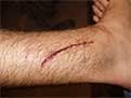
A laceration to the leg

A lacerated knee

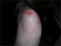
An infected puncture wound to the bottom of the forefoot.

A puncture wound from playing darts.

Pathophysiology

To heal a wound, the body undertakes a series of actions collectively known as the wound healing process.

Management

Wound, sewn with four stitches

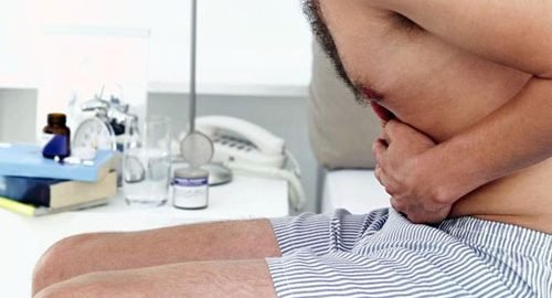
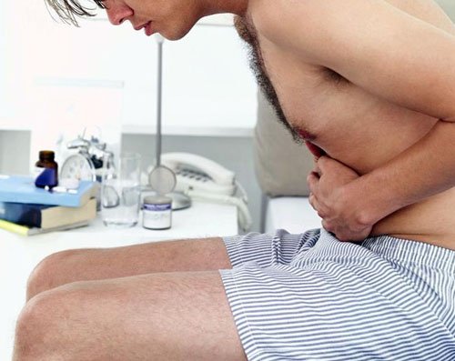
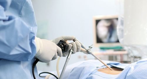
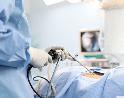
The overall wound treatment in Delhi depends on the type, cause, and depth of the wound as well as whether or not other structures beyond the skin (dermis) are involved. Treatment of recent lacerations involves examining, cleaning, and closing the wound. Minor wounds, like bruises, will heal on their own, with skin discoloration usually disappearing in 1–2 weeks. Abrasions, which are wounds with intact skin (non-penetration through dermis to subcutaneous fat), usually require no active treatment except keeping the area clean, initially with soap and water. Puncture wounds may be prone to infection depending on the depth of penetration. The entry of puncture wound is left open to allow for bacteria or debris to be removed from inside.

Cleaning

Evidence to support the cleaning of wounds before closure is poor. For simple lacerations, cleaning can be accomplished using a number of different solutions, including tap water and sterile saline solution. Infection rates may be lower with the use of tap water in regions where water quality is high. Cleaning of a wound is also known as wound toilet.
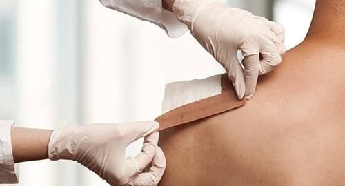
Closure

If a person presents to a healthcare center within 6 hours of a laceration they are typically closed immediately after evaluating and cleaning the wound. After this point in time, however, there is a theoretical concern of increased risks of infection if closed immediately. Thus some healthcare providers may delay closure while others may be willing to immediately close up to 24 hours after the injury. A single study has found that using clean non sterile gloves is equivalent to using sterile gloves during wound closure.

If closure of a wound is decided upon a number of techniques can be used. These include bandages, a cyanoacrylate glue, staples, and sutures. Absorbable sutures have the benefit over non absorbable sutures of not requiring removal. They are often preferred in children. Buffering the pH of lidocaine makes the freezing less painful.

Adhesive glue and sutures have comparable cosmetic outcomes for minor lacerations 5 cm in adults and children. The use of adhesive glue involves considerably less time for the doctor and less pain for the person with the cut. The wound opens at a slightly higher rate but there is less redness. The risk for infections (1.1%) is the same for both. Adhesive glue should not be used in areas of high tension or repetitive movements, such as joints or the posterior trunk.

Dressings

The effectiveness of dressings and creams containing silver to prevent infection or improve healing is not currently supported by evidence.

Imaging

A wound may be recorded for follow-up and observing progress of healing with different techniques which consist of, but not limited to:

  • Photographs, with subsequent area quantification using computer processing
  • Wound tracings on acetate sheets
  • Kundin wound gauge


Complications

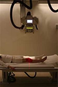
The patient has a deep wound at the knee, and radiography is used to ensure there is no hidden bone fractures.

Bacterial infection of wound can impede the healing process and lead to life threatening complications. Scientists at Sheffield University have identified a way of using light to rapidly detect the presence of bacteria. They are developing a portable kit in which specially designed molecules emit a light signal when bound to bacteria. Current laboratory-based detection of bacteria can take hours or even days.

Workup

Individuals who have wounds that are not healing should be investigated to find the causes. Many microbiological agents can be responsible for this. The basic workup includes evaluating the wound, its extent and severity. Cultures are usually obtained both from the wound site and blood. X rays are obtained and a tetanus shot may be administered if there is any doubt about prior vaccination.

Chronic

Non-healing wounds of the diabetic foot are considered one of the most significant complications of diabetes, representing a major worldwide medical, social, and economic burden that greatly affects patient quality of life. Almost 30 million Indians-one in every 10-are diabetic and the disease is causing widespread disability and death at an epidemic pace. Of those with diabetes, 6.5 million are estimated to suffer with chronic or non-healing wounds. Associated with inadequate circulation, poorly functioning veins, and immobility, non-healing wounds occur most frequently in the elderly and in people with diabetes-populations that are sharply rising as the nation ages and chronic diseases increase and thus it requires proper diabetic foot treatment in Delhi.

Although diabetes can ravage the body in many ways, non-healing ulcers on the feet and lower legs are common outward manifestations of the disease. Also, diabetics often suffer from nerve damage in their feet and legs, allowing small wounds or irritations to develop without awareness. Given the abnormalities of the microvasculature and other side effects of diabetes, these wounds take a long time to heal and require a specialized treatment approach for proper healing.

As many as 25% of diabetic patients will eventually develop foot ulcers, and recurrence within five years is 70%. If not aggressively treated, these wounds can lead to amputations. It is estimated that every 30 seconds a lower limb is amputated somewhere in the world because of a diabetic wound. Amputation often triggers a downward spiral of declining quality of life, frequently leading to disability and death. In fact, only about one third of diabetic amputees will live more than five years, a survival rate equivalent to that of many cancers.

Many of these lower extremity amputations can be prevented through an interdisciplinary approach to treatment involving a variety of advanced therapies and techniques, such as debridement, hyperbaric oxygen treatment therapy, dressing selection, special shoes, and patient education. When wounds persist, a specialized approach is required for healing.
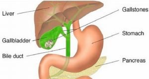
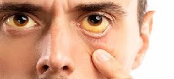
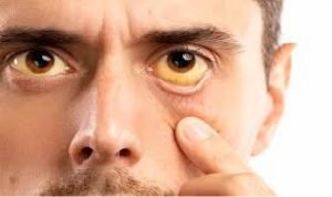