Category Archives: Service

Anal Fissures

anal-fissure-treatment-in-Delhi
The anal fissure is a longitudinal tear that appears in the most distal portion of the anal canal and may require medical or surgical anal fissure treatment in Delhi. Its most frequent location is the posterior midline (90-98%), another less common location is the anterior midline (12% of those that appear in women and 7% in men). It has an equal incidence in both sexes and is more frequent in the middle age of life. Most are of unknown origin. The most likely explanation is an acute traumatism of the anal canal, defecation (large, hard stools) and rarely due to the explosive expulsion of liquid stools. The fact that the posterior wall of both the subendothelial space and the sphincter are less vascularized makes them more vulnerable to the location of the fissures. The transition to chronicity is due both to sphincter hypertonia and ischemia.

The presence of multiple fissures or in places other than those mentioned requires us to rule out diseases such as ulcerative colitis, Crohn’s disease, tuberculosis, syphilis, immunodeficiency syndrome … (it must be taken into account that more than half of the fissures secondary to intestinal inflammatory diseases occur in the posterior midline and are painful).

Symptoms

The main symptom of a fissure is a pain. It is an intense pain that is triggered by defecation, lasting in a variable way, from a few minutes to hours. There may also be bleeding, itching or itching and inflammation of the base of the fissure, which is often confused with hemorrhoid (“sentinel hemorrhoid”) and causes misdiagnosis if a laparoscopic surgeon in Delhi is not consulted.

The formation of an anal fissure results in the establishment of a “vicious circle” that hinders or prevents its healing: the existence of the fissure causes the defecation to produce pain, the pain causes a hypertonia or reflex spasm of the internal anal sphincter (responsible for the pain after defecation); hypertonia of the sphincter produces an alteration of the vascularization of the anal region that hinders the healing of the fissure, thus favoring the persistence of the “vicious circle”

Inspection

It should be done with great care because given the hypertonia of the canal it is very painful. The patient is placed in the left lateral decubitus position (Sims position), the anal margins are separated and the patient is asked to perform a Valsalva maneuver (it is often necessary to previously apply a topical anesthetic or oral analgesics). Rectal examination is contraindicated due to the risk of vasovagal syncope and even cardiac arrest; if it could be done, it would show sphincter hypertonia. Anoscopy and rectoscopy are only necessary when a secondary fissure is suspected.

Anal Fissure Treatment in Delhi

The therapeutic attitude will be different before an acute form or if signs of chronicity already exist (deep fissure, sentinel hemorrhoids, etc).

In the acute phase is when medical treatment has more chances of success. This consists of :

  • Abundant intake of fluids
  • Diet rich in fiber.
  • Additives that contain fiber such as Plantago (Plantaben, cenat, etc.)
  • Seat baths with warm-hot water.
  • Topical ointments
  • In patients with a high risk of incontinence: botulinum toxin type A

Can the fissure be operated?

It must be taken into account that medical anal fissure treatment in Delhi should always be attempted, even in chronic cases, where the percentage of success is smaller. The success rate of medical treatment is 60%.

The surgical choice of anal fissure treatment in Delhi is the internal lateral sphincterotomy. The intervention consists of the controlled section of the internal anal sphincter that eliminates the hypertonia of the same, disappearing the pain and getting the healing of the fissure in a few days. The operation is carried out by sedation + local anesthesia or spinal anesthesia under outpatient surgery.

Other techniques such as anal dilation have become obsolete or limited to very specific cases, especially after the demonstration of unsuspected sphincter injuries after anal dissemination in endoanal ultrasound studies.

Anal Fistula

Anal-Fistula-Treatment-in-Delhi
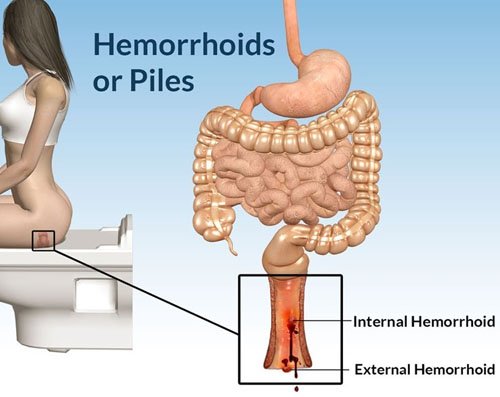
Anal fistula treatment in Delhi is done for a very painful fistula appeared near the anus. A fistula is an abnormal connection (formation of a channel) between two parts, which occurs inside the body. It can be formed between two organs, between the intestine and another structure, between two blood vessels, between an artery and a vein, and so on. In the specific case of an anal fistula, a tube-shaped duct forms under the skin surrounding the anus. It consists, therefore, of an external orifice (in the skin of the anus) and an internal one (inside the anus or rectum) existing communication between both, although there are complex fistulous trajectories that do not communicate these orifices. They are usually the consequence of an anal abscess that has evolved in most cases.

An anal fistula is a relatively common disease, especially in adults, although it can occur in children if it is associated with intestinal inflammatory diseases such as Crohn’s disease. It appears more frequently in males than in females. Its prevalence is estimated at around 30%, with a peak of age around 40 years.

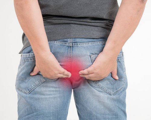
The clinical picture of this disorder can be practically asymptomatic if it is in a zone somewhat distant from the anus, with mild discomfort of itching or stinging, or manifest as a sharp pain, very intense and cutting nature that occurs at the time of passage of the stool in the area during the deposition, and that may remain for several hours after the same. Occasionally there may be slight bleeding and small amounts of pus.

Causes

In general, an anal fistula can be caused by the presence of some type of wound, an injury produced during surgery, infection or inflammation. The obstruction of a gland is the situation that is most commonly associated with the formation of fistulas.

The accumulation of any liquid in a body cavity favors the appearance of infections so that if the secretion product of the anal glands does not find an exit route it is very likely that the enteric bacteria (bacterial flora found in the intestine of healthy individuals) provoke an infection in a short time. Immediately, pus begins to form and accumulate, forming an abscess (very localized lesion, characterized by the accumulation of pus inside it) that usually ends up draining at some point in the anus. This is the cause of approximately 80-90% of cases.

The abscesses in this area may also be formed by the infection of an anal fissure (small break in the mucus covering the anus) or a sexually transmitted infection contact.

Symptoms

The most frequent symptoms for which a person with anal fistula consults the laparoscopic surgeon in Delhi are a pain in the area of ​​the anus and the presence of pus or drainage of some malodorous secretion.

On many occasions, these individuals have previously consulted for infections of the area or true abscesses. Or have presented pain and pus secretion for a long time, but have not consulted because the symptoms were intermittent or modesty.

The pain is variable in intensity, being able to accentuate when defecating when sitting down, or on other occasions when coughing or sneezing due to the increase of pressure generated by these situations. As a difference from other pathologies in this region, the pain generated by abscesses or fissures is usually more intense, besides having a red and hard lump in the case of abscesses.

You can see the external orifices of the fistula, sometimes several, through which it expels purulent, malodorous, sometimes even fecaloid (vomit of fecal material) content. This secretion usually causes irritation of the perianal skin, presenting itching, stinging and redness of the area.

When the laparoscopic surgeon in Delhi digitally explores the anal and rectal area, it can identify which type of fistula is attending the classification described at the beginning, depending on the relationship with the external and internal sphincter, as well as estimate the length and complexity of the fistulous tract.

Diagnose

A physical examination will be carried out in the first instance to the patient who comes to consult with discomforts that may make us suspect the presence of an anal fistula. The doctor will look for the presence of abscesses and openings in the skin that shows the existence of an anal fistula. Normally a zone of reddened skin will be observed and painful to the touch. If it is found, it will try to define its route and depth with the help of a probe.

The presence of fistulas is not always evident since the lesion does not have to be on the surface of the skin. For this reason, it is sometimes necessary to perform a digital rectal examination and observe the anal canal and rectum with the help of an anoscope; This instrument is a short and rigid tube with a light at the end, which allows to observe the inside of the cavity and take samples.

An important point in the diagnosis of an anal fistula is to find its origin. Since there are other pathologies that can lead to the appearance of abscesses and fistulas, to determine if the underlying cause is, for example, some type of cancer, Crohn’s disease or other pathology, a sigmoidoscope examination will be performed (sigmoidoscopy). This instrument is a long and flexible tube that allows you to see the large intestine up to 60 cm inward from the anus.

On other occasions anal or endoanal ultrasound is useful by means of a rotating probe to determine the path, route, presence of other secondary paths and collections of pus.

At present, magnetic resonance imaging is also available to obtain information on the characteristics of the fistula or fistulas and their complexity, as well as the presence of adjacent complications. TAC can also be used if contrast is added to supplement the study.

Anal Fistula Treatment in Delhi

The only effective anal fistula treatment in Delhi for those patients who suffer it is surgical intervention. By means of this procedure, it is intended to definitively eliminate the fistula and correct the alterations that may have appeared as a result of it, without compromising the anal continence. The disappearance of the fistula implies the disappearance of the associated painful discomfort, inflammation, suppuration …

Depending on the type of fistula, its path, depth, etc., different types of intervention may be performed.

  • Fistulotomy: it consists of the opening and emptying of the fistulous tract, trying to ensure healing as efficiently and quickly as possible.
  • Fistulectomy: this anal fistula treatment in Delhi involves the complete removal of the fistulous tract. The drawback with respect to the prior art is that it gives rise to larger wounds and, therefore, the healing is slower and more expensive.
  • Sedal or seton: there are three variants of this technique depending on the objective of the intervention.
    • Cutting seton: the objective of this modality is the elimination of the fistula. It is done by passing through its surgical silk that is tightened gradually so that the silk section the sphincter but at the same time this has enough time to go scarring.
    • Drainage seton: performed to drain pus or debris from the fistula; in this way, the formation of a new abscess will be avoided. This silk will be placed through the fistula, and once the drainage process is complete it allows surgical repair even in the absence of infection.
    • Seton guide: it is placed in the same way as the previous one. Its function is to keep the fistulous tract patent until the patient can be definitively operated on. This method is usually used in patients with recurrent perianal abscesses, in which the point where the abscess was found cannot be identified since there is no sequela of the fistulous tract.

In the case of anal fistulas related to Crohn’s disease, the initial anal fistula treatment in Delhi is not surgery, but the management with metronidazole antibiotics, to avoid the operating theater, as well as to reinforce the systemic treatment of the disease with azathioprine-type immunosuppressants or agents. biological agents such as adalimumab or infliximab. The techniques described above are resorted to if they fail.

For pain relief prior to and after the intervention, analgesics and common anti-inflammatories will be useful, and it is important to achieve non-painful defecation through proper hydration and an adequate supply of fiber to the diet.

Prevention

The prevention of anal fistulas implies the prevention of all those factors that can trigger them:

  • Fissures. In adults, it is common to have fissures due to constipation or prolonged diarrhea. It is important to have a balanced diet, rich in fiber, to ensure proper intestinal transit, and avoid foods that may cause irritation.
  • The appearance of fissures and perianal abscesses is also common in babies. To prevent them it is advisable to change diapers often and clean it properly.

In addition, usual measures should be taken into account such as:

  • Clean after defecation with soft materials or water.
  • Avoid a sedentary lifestyle; the physical activity strengthens the immune system.
  • Use prophylactic measures when having sex.

It is important to consult a laparoscopic surgeon in Delhi when you perceive anal pain and spotting of underwear or non-fecal material with a bad smell, to avoid that in the case of abscesses, these evolve to the formation of the fistula.

In addition to these basic measures, you can try some homeopathic practices, not as a treatment, but as a prevention or as a way to acquire healthier habits, in order to avoid the reappearance of injuries already treated. With regard to food, there are for example certain foods that are attributed anti-inflammatory properties (ginger, turmeric, vegetables, and fruits), refined products such as sugar, fried foods, and other processed foods have the opposite effect, therefore, It may be beneficial to reduce your consumption.

Hemorrhoids (Piles)

piles-treatment-in-delhi
Hemorrhoidal disease is one of the most common diseases in humans. More than half of the population has symptoms of hemorrhoids. Millions of people have hemorrhoids (piles). A common patient suffers “in silence” for a long time before consulting a laparoscopic surgeon in Delhi. Currently, there is piles treatment in Delhi that allows quick recovery and with little pain and discomfort.

What are they?

Hemorrhoids are normal anatomical formations. Each individual is born with hemorrhoids. When they become symptomatic, we refer to a patient as a carrier of hemorrhoidal disease.

How often do they have?

More than 60% of the population over the age of 50 will suffer from hemorrhoid symptoms.

What are they for?

Hemorrhoids are important for maintaining “fine” continence. Acting as “anal pads, they ensure a perfect closure of the anal canal.

Why do they generate problems?

Most symptoms are caused by the prolapse (slippage) of the anorectal mucosa in and out of the anal canal. The prolapse of the mucosa and internal hemorrhoids in the anal canal causes sphincter pressure on these structures to determine an alteration of blood flow, which predisposes to “thrombosis” (clots within the blood vessels).

What factors predispose to the appearance of hemorrhoids?

  • Diarrhea and constipation.
  • Difficulty evacuation and abuse of laxatives.
  • Prolonged efforts.
  • Sedentary life.
  • Certain sports such as cycling or horse riding.
  • Pregnancy and childbirth
  • Circulatory disorders
  • Alcohol abuse.
  • Spicy food abuse.
  • Excess coffee.

What symptoms do they produce?

External hemorrhoids generally do not bleed. They can thrombosis (clots inside the blood vessels) and cause great pain. Although external hemorrhoids can rarely become necrotic and cause a serious complication, most thrombosed hemorrhoids resolve spontaneously. Subsequently, redundant areas of skin may remain (external plicomas). These can cause itching and difficulty in performing a proper toilet.

Symptoms generally include increased local volume and inflammation, pain in relation to defecation or the presence of hemorrhoidal mass and defecatory and/or spontaneous anal bleeding, which may be scarce or lead to chronic anemia. In some patients, severe bleeding may occur.

How is the diagnosis of hemorrhoids made?

The diagnosis is made with a proctological examination performed by a laparoscopic surgeon in Delhi. Although the hemorrhoidal masses are clearly visualized, a rectal endoscopy should be performed to rule out other lesions. The presence of underlying diseases such as inflammatory bowel diseases, tumors or colorectal polyps should always be ruled out.

Can hemorrhoids become cancer?

No. But the same symptoms produced by hemorrhoids can be due to a tumor, or other serious diseases, especially bleeding.

How are piles treatment in Delhi done?

Pils treatment in Vikaspuri, Janakpuri, Dwarka includes:

  • Over-the-counter corticosteroid creams (for example, cortisone) to help decrease pain and swelling.
  • Hemorrhoid creams that contain lidocaine to help reduce pain.
  • Stool softeners help reduce effort and constipation.

Things that can be done to reduce itching include:

  • Apply witch hazel to the area with cotton spots.
  • Wear cotton underwear.
  • Avoid toilet paper with perfumes or colors; use baby wipes instead.
  • Try not to scratch the area.

Depending on the grade and symptoms, piles treatment in Delhi can be done with hygienic-dietary measures, non-surgical procedures or surgery.

The initial piles treatment in West Delhi, Vikaspuri, Janakpuri, Dwarka consists of conservative measures, such as a diet rich in fiber, fiber supplements, mild laxatives, local hygiene measures, etc. If symptoms persist, it is recommended to perform procedures or surgery according to the degree of hemorrhoids.

Hemorroidopexy with mechanical suture

This is the first method that was an alternative to the removal of hemorrhoids. This method, known as the Longo or PPH technique, involves performing a hemorrhoidopexy. The mucosal prolapse is corrected by removing a part of the rectal canal, replacing the hemorrhoids in the original position. A mechanical self-stapler is used that removes the remaining mucosa and, at the same time, sutures the remainder. Postoperative pain is markedly reduced because there are no open wounds. This piles treatment in Janakpuri indicated for hemorrhoids of 2nd, 3rd and 4th grade.

bariatric-surgery-in-delhi

Bariatric Surgery

bariatric-surgery-in-delhiObesity is today one of the main concerns of the World Health Organization (WHO). Already considered as a pandemic, overweight and obesity in India have alarming rates, leading the rankings internationally. Every year thousands of people opt to go for bariatric surgery in Delhi to curb obesity. According to the report, 74% of Indians are overweight and 86% are sedentary, while 32% suffer from obesity, and 3% morbidly obese. In the world, 44% of global cases of diabetes, 23% of ischemic heart disease and a significant percentage of certain cancers are attributable to overweight and obesity, ”says bariatric surgeon in Delhi.

Dr. Mohit Jain, who is a famous bariatric surgeon in Delhi, points out that there are three types of patients who are more recurrent in this type of procedure: the obese patient, the obese diabetics, and the patients who have gained weight again after bariatric surgery. He also points out that women are more recurrent patients than men, and that more teenagers and young people are treated every day, although the most frequent range is between forty and fifty years.

What is bariatric surgery?

It is a surgical intervention to treat morbid obesity and obesity disease, understanding obesity as an excessive overweight that puts health and quality of life at risk, corresponding to having a body mass index (BMI) or relationship between weight and height, greater than forty. Obesity is a disease because it has all the properties of the disease. That is, it compromises the health of the organism, and decreases the years and quality of life. The bariatric surgery in Delhi is currently the only long-term therapeutic effective means of inducing a satisfactory weight loss and definitive and in the control, or in the resolution, the dell ‘complications obesity.

The goal of bariatric surgery in Delhi is therefore not only the reduction and maintenance of body weight but also the prevention and treatment of complications, and more generally the improvement of the quality of life.

Types of Bariatric Surgeries

  • Restrictive: This type of procedure changes the size of the stomach to reduce food intake in large quantities, keeping the digestive system functions intact
  • Malabsorptive: These change the way in which the digestive system carries out its processes, change the path of food, preventing them from passing through a part of the stomach and small intestine that absorb calories.

Within these two types, which can be combined, there are also different techniques to perform the interventions, according to the bariatric surgeon in Delhi. 70% of the people attended the clinic correspond to Gastric Sleeve, while the Bypass adds thirty percent of the care.

Interventions of the Bariatric Surgeon in Delhi:

  • Sleeve gastrectomy
  • Gastric bypass
  • Gastric bandage
  • Endoscopic intragastric balloon

They are based on the reduction of the gastric volume, which determines an early sense of satiety with a very reduced food intake, which leads to the loss of body weight. These interventions involve a lower physiological impact, since they do not modify the processes of digestion, but result in a lower reduction of overweight.

The Gastric Sleeve is an operation where the volume of the stomach is reduced by 80%, leaving a non-distensible tube, that is to say rigid, which limits the amount of food that is eaten, producing an important weight loss. This technique of bariatric surgery in Delhi has the particularity of eliminating Ghrelin, a hormone that is located in the stomach and that stimulates the appetite. “It is an operation that is associated with a prolonged period in which we have little hunger, which lasts between eight months and one year after the intervention,” says bariatric surgeon in Delhi. This procedure is recommended for cases of obesity of up to fifty kilos of overweight.

On the other hand, the Gastric Bypass is recommended for cases of obesity where overweight exceeds fifty kilos, as well as for diabetic patients, due to their metabolic effects, whether obese or not. The Bypass represents 30% of the care. The intervention consists of dividing the stomach into two parts, leaving a blocked part that does not receive food. In addition, food is less assimilated by a fraction of the stomach, having a mixed, restrictive and malabsorptive effect.

Indications for bariatric surgery in Delhi

  • Aged between 18 and 65;
  • Motivated and collaborative patient;
  • History of the obesity dating back at least 5 years;
  • Obesity with  BMI  greater than 40 or with a BMI  between 35 and 40 with the presence of complications related to obesity;
  • Failure of previous non-surgical therapies, correctly followed for at least a year;
  • Absence of contraindications to surgery.

Diagnostic Laparoscopy

Diagnostic Laparoscopy in Delhi
Diagnostic Laparoscopy Patient Information

Patients may be referred to surgeons because of an undiagnosed abdominal problem. If your surgeon in Delhi has recommended a diagnostic laparoscopy, this page will:

  • help you understand what laparoscopy is,
  • describe how laparoscopy helps to find out what the problem is,
  • explain what complications can occur with the procedure.


What is Diagnostic Laparoscopy?

A laparoscope is a telescope designed for medical use. It is connected to a high-intensity light and a high-resolution television camera so that the surgeon can see what is happening inside of you. The laparoscope is put into the abdominal cavity through a hollow tube and the image of the inside of your abdomen is seen on the television screen. In most cases, this procedure (operation) will be able to diagnose or help discover what the abdominal problem is.

Why Is Diagnostic Laparoscopy Performed?

1. Abdominal pain.
Laparoscopy has a role in the diagnosis of both acute and chronic abdominal pain. There are many causes of abdominal pain. Some of these causes include appendicitis, adhesions or intra-abdominal scar tissue, pelvic infections, endometriosis, abdominal bleeding and, less frequently, cancer. It is used in patients with irritable bowel disease to exclude other causes of abdominal pain. Laparoscopic surgeon in Delhi can often diagnose the cause of the abdominal pain and, during the same procedure, correct the problem.

2. Abdominal mass.
A patient may have a lump (mass or tumor), which can be felt by the doctor, the patient, or seen on an X-ray. Most masses require a definitive diagnosis before appropriate therapy or treatment can be recommended. Laparoscopy is one of the techniques available to your physician to look directly at the mass and obtain tissue to discover the diagnosis.

3. Ascites.
The presence of fluid in the abdominal cavity is called ascites. Sometimes the cause of this fluid accumulation cannot be found without looking into the abdominal cavity, which can often be accomplished with laparoscopy.

4. Liver disease.
Non-invasive X-ray imaging techniques (sonogram, CT scan, and MRI) may discover a mass inside or on the surface of the liver. If the non-invasive X-ray cannot give your physician enough information, a liver biopsy may be needed to establish the diagnosis. Diagnostic laparoscopy is one of the safest and most accurate ways to obtain tissue for diagnosis. In other words, it is an accurate way to collect a biopsy to sample the liver or mass without actually opening the abdomen.

5. “Second look” procedure or cancer staging.
Your doctor may need information regarding the status of a previously treated disease, such as cancer. This may occur after treatment with some forms of chemotherapy or before more chemotherapy is started. Also, information may be provided by diagnostic laparoscopy before planning a formal exploration of the abdomen, chemotherapy or radiation therapy.

6. Other.
There are other reasons to undergo a diagnostic laparoscopy, which cannot all be listed here. This should be reviewed and discussed with your surgeon.

What Tests Are Necessary Before Laparoscopy?

Ultrasound may be ordered by your doctor as a non-invasive diagnostic test. In many cases, information is provided which will allow your surgeon to have a better understanding of the problem inside your abdomen. This test is not painful, is very safe, and can improve the effectiveness of the diagnostic laparoscopy.

CT Scan is an X-ray that uses computers to visualize the intra-abdominal contents. In certain circumstances, it is accurate in making the diagnosis of abdominal disease. It will allow your surgeon to have a “road map” of the inside of your abdomen. A radiologist may use a CT scan to place a needle inside your abdomen. This is known as a CT guided needle biopsy. This will often be done before a diagnostic laparoscopy to decide if laparoscopy is appropriate for your condition.

MRI (magnetic resonance imaging) uses magnets, X-rays, and computers to view the inside of the abdominal cavity. It is not required for most abdominal problems but may be necessary for some.

Routine blood test analysis, urinalysis, and possible chest X-ray or electrocardiogram may be needed before diagnostic laparoscopy. Your physician will decide which tests are necessary and will review the results of those tests, which have already been performed.

What Type of Anesthesia Is Used?

Diagnostic laparoscopy is performed either under local anesthesia with sedation or with general anesthesia. With your help, your surgeon and an anesthesiologist will decide on a method of anesthesia to perform safe and successful laparoscopic surgery in Delhi.

LOCAL anesthesia can be injected into the skin of the abdominal wall to completely numb the area and allow safe placement of a laparoscope. Most patients feel a short-lived “bee sting” that lasts a second or two. Small doses of intravenous sedation are given at the same time allowing the patient to experience what is known as “twilight” sleep in which patients are arousable but asleep. Once an adequate depth of sleep is reached and local anesthesia administered, gas is placed into the abdominal cavity. This is called a pneumoperitoneum. The patient may experience a bloated feeling. The gas is removed at the end of the operation. The two most common gases used are a nitrous oxide (“laughing gas”) or carbon dioxide. There is very little risk of ill-effects of the gas.

GENERAL anesthesia is given to those patients who are not candidates for “twilight” sleep or who want to be completely asleep. General anesthesia may be preferable in patients who are young, who cannot lie still on the operating table, or have a medical condition that is safer to perform in this manner. Some patients end up having general anesthesia even though they prefer local anesthesia with sedation, as the appropriate anesthesia for laparoscopy differs from patient to patient.

What Preparation Is Required?

  • After your surgeon reviews with you the potential risks and benefits of the operation, you will need to provide written consent for surgery.
  • It is acceptable to shower the night before or the morning of the operation.
  • Most diagnostic laparoscopy procedures are performed as an outpatient; meaning you will go home the same day the procedure was performed.
  • You should have nothing to eat or drink for six to eight hours before the procedure.
  • Standard blood, urine, or X-ray testing may be required before your operative procedure. This will depend on your age and medical conditions.
  • It is acceptable to shower the night before or the morning of the operation. Report to the hospital at the correct time, which is usually 1-2 hours earlier than your scheduled surgery.
  • If you take medication on a daily basis, discuss this with your surgeon prior to surgery as you may need to take some or all of the medication on the day of surgery with a sip of water. If you take aspirin, Vitamin E, blood thinners or arthritis medication, discuss this with your surgeon so they can be stopped at the proper time before your surgery.
  • You will need to ask your surgeon or his/her office staff what specifically is required in preparation for your surgery.


You will most likely be sedated during the procedure and an arrangement to have someone drive you home afterward is imperative. Sedatives will affect your judgment and reflexes for the rest of the day. You should not drive or operate machinery until the next day.

What Can Be Expected During Diagnostic Laparoscopy?

  • The surgery is performed under anesthesia (see above), so that the patient will not feel pain during the procedure.
  • A cannula (a narrow tube-like instrument) is placed into the abdominal cavity in the upper abdomen or flank just below the ribs.
  • A laparoscope (a tiny telescope) connected to a special camera is inserted through the cannula. This gives the surgeon a magnified view of the patient’s internal organs on a television screen.
  • Other cannulas are inserted which allow your surgeon to see the internal organs and make a decision on the proper diagnosis or treatment
  • After the surgeon completes the operation, the small incisions are closed with absorbable sutures or with surgical tapes.


What Should I Expect After the Operation?

Following the operation, you will be transferred to the recovery room, where you will be monitored carefully until all the sedatives and anesthetics have worn off. Even though you may feel fully awake, the effects of any anesthetic may persist for several hours. Once you are able to walk and get out of bed unassisted, you may be discharged. Because the effects of anesthesia can linger for many hours, it is necessary to have someone accompany you to the office or hospital and drive you home after the procedure.

You can expect some soreness around any incision site; this is normal. Your pain should improve daily even though you may need to take a pain reliever. Your surgeon will instruct you on the use of pain relievers and may give you a prescription for pain medication.

Most patients are able to shower the day after surgery and begin all normal activities within a week. Your surgeon can answer any specific restrictions that apply to you.

You should call and schedule a follow-up appointment within two weeks after your procedure.

What Complications Can Occur?

Any procedure may have complications associated with it. The most frequent complications of any operation are bleeding and infection. There is a small risk of other complications that include, but are not limited to, injury to the abdominal organs, intestines, urinary bladder or blood vessels. If you suffer from ascites, this ascites may leak from one of the operative sites, temporarily, before stopping.

In a small number of patients, the laparoscopic method cannot be performed. The decision to perform the open procedure is a judgment decision made by your surgeon either before or during the actual operation. When the surgeon feels that it is safest to convert the laparoscopic procedure to an open one, this is not a complication, but rather sound surgical judgment. The decision to convert to an open procedure is strictly based on patient safety

When To Call Your Doctor

Be sure to call your surgeon or physician if you develop any of the following:

  • fever above 101 degrees F (39 C)
  • drainage from or redness any of your incisions
  • continued nausea or vomiting
  • increasing abdominal swelling
  • bleeding
  • chills
  • persistent cough or shortness of breath
  • inability to urinate
  • pain not controlled by medication

Laparoscopic Appendectomy

appendix-surgery-in-delhi
The appendix produces a bacteria-destroying protein called immunoglobulin, which helps fight infection of the organism. However, its function is not essential. People who have had appendix surgery in Delhi do not have an increased risk of infection. Other organs of the body assume this function when the appendix is ​​removed.

What is a laparoscopic appendectomy?

Appendicitis is one of the most common surgical problems. An appendix removal in Delhi is performed at one time in every one thousand people. Treatment requires an operation to remove the infected appendix. Traditionally, the appendix is removed through an incision in the lower right abdominal wall.

In most laparoscopic appendectomies, a laparoscopic surgeon in Delhi operates through three small incisions (each measuring approx. 6-12.5 mm) while viewing an enlarged image of the patient’s internal organs on a television screen. In some cases, one of the small openings can be extended to measure 5 to 7.5 cm in order to complete the procedure.

Advantages of laparoscopic appendectomy

The results may vary depending on the procedure used and the general condition of the patient. The common advantages are:

  • Less postoperative pain
  • May shorten hospital stay
  • It can result in a faster return of bowel function
  • Faster return to normal activity
  • Better cosmetic results

Are you a candidate for a laparoscopic appendectomy?

While laparoscopic appendectomy has many benefits, it may not be appropriate for some patients. Unperforated appendicitis that is diagnosed early can usually be removed laparoscopically. Laparoscopic appendix treatment in Delhi is more difficult to perform if there is a severe infection or if the appendix has perforated. It is possible that in such patients it will be necessary to perform an open-air procedure in which a larger incision is used in order to safely remove the infected appendix.

How is laparoscopic appendectomy carried out?

The words “laparoscopic” and “open pit” appendectomy describe the techniques a surgeon uses to access the internal surgical field. For the most part, laparoscopic appendix surgery in Delhi begins in the same way. Through the use of a cannula (a narrow and tubular instrument), the surgeon accesses the abdomen.

A laparoscope (a small telescope connected to a camcorder) is inserted through a cannula, which offers the surgeon in Delhi an enlarged view of the patient’s internal organs on a television screen. Several additional cannulas are inserted to allow the surgeon to work inside and remove the appendix. The entire procedure can be completed through the cannulas or by lengthening one of the small incisions for the cannulas. A drain may be placed during the procedure. It will be removed before you leave the hospital.

What happens if the operation cannot be performed or if it cannot be completed by the laparoscopic route?

In a small number of patients, the laparoscopic method is not feasible because of the inability to visualize or manipulate the organs effectively. When the surgeon feels that it is safest to convert the laparoscopic procedure into an open pit, it is not a complication, but a sensible surgical decision. Among the factors that may increase the possibility of the procedure becoming an “open pit” may include:

  • Severe infection and / or an abscess
  • A perforated appendix
  • Obesity
  • History of previous abdominal surgery that produced dense scar tissue
  • Inability to visualize organs
  • Bleeding problems during the operation

The decision to perform the procedure outdoors is determined at the discretion of your surgeon either before or during the operation itself. The decision to convert to an open procedure is based exclusively on patient safety.

What is to be expected after the appendix surgery in Delhi?

After the operation, it is important that you follow your doctor’s instructions. While some people feel better after a few days, remember that your body needs time to heal. You are encouraged to get out of bed the day after surgery and to walk. This will help decrease your muscle aches and also the risk of clot formation in the legs.

You may be able to resume most of your normal activities after one or two weeks. These activities include: taking showers, driving your car, climbing stairs, working and having sex. If your pain is prolonged or if you are not relieved by prescription pain relievers, you should inform your laparoscopic surgeon in Delhi. Call and ask for a medical control shift before the two weeks following your operation.

What complications can occur?

As with any operation, there are risks, including the risk of complications. However, the risk of any of these complications occurring is not greater than if the operation was performed using the open-pit technique.

  • Bleeding
  • Infection
  • Removal of a normal appendix
  • A loss at the edge of the colon where the appendix was removed
  • Injury to adjacent organs such as the small intestine, ureter or bladder.
  • A blood clot to the lungs

It is important that you recognize the early signs of possible complications. Contact your surgeon if you notice severe abdominal pain, fever, chills or rectal bleeding.

When should you call your doctor?

Be sure to call your doctor or surgeon if any of the following symptoms occur:

  • Fever above 101ºF (39ºC) that does not yield
  • Bleeding
  • Abdominal swelling that is increasing
  • Pain that is not relieved by taking your medications
  • Nausea or persistent vomiting
  • Shaking chills
  • Persistent cough or shortness of breath
  • Purulent drainage (pus) of any incision
  • Redness around any of your incisions that worsens or enlarges
  • Inability to eat or drink liquids

Laparoscopic Colon Resection

colon-surgery-in-delhi
About Conventional Colon Surgery…

Each year, more than 600,000 surgical procedures are performed in India to treat a number of colon diseases. Although colon surgery in Delhi is not always a cure, it is often the best way to stop the spread of disease and alleviate pain and discomfort.

Patients undergoing colon surgery in Delhi often face a long and difficult recovery because the traditional “open” procedures are highly invasive. In most cases, surgeons are required to make a long incision. Surgery results in an average hospital stay of a week or more and usually 6 weeks of recovery.

WHAT IS THE COLON?

The colon is the large intestine; it is the lower part of your digestive tract. The intestine is a long, tubular organ consisting of the small intestine, the colon (large intestine) and the rectum, which is the last part of the colon. After food is swallowed, it begins to be digested in the stomach and then empties into the small intestine, where the nutritional part of the food is absorbed. The remaining waste moves through the colon to the rectum and is expelled from the body. The colon and rectum absorb water and hold the waste until you are ready to expel it.

WHAT IS LAPAROSCOPIC COLON SURGERY IN DELHI?

A technique known as minimally invasive laparoscopic colon surgery allows laparoscopic surgeon in Delhi to perform many common colon procedures through small incisions. Depending on the type of procedure, patients may leave the hospital in a few days and return to normal activities more quickly than patients recovering from open surgery.
In most laparoscopic colon resections, surgeons operate through 4 or 5 small openings (each about a quarter-inch) while watching an enlarged image of the patient’s internal organs on a television monitor. In some cases, one of the small openings may be lengthened to 2 or 3 inches to complete the procedure.

WHAT ARE THE ADVANTAGES OF LAPAROSCOPIC COLON SURGERY IN DELHI

Results may vary depending upon the type of procedure and patient’s overall condition. Common advantages are:

  • Less postoperative pain
  • May shorten hospital stay
  • May result in a faster return to solid-food diet
  • May result in a quicker return of bowel function
  • Quicker return to normal activity
  • Improved cosmetic results


ARE YOU A CANDIDATE FOR LAPAROSCOPIC COLON RESECTION?

Although laparoscopic colon surgery in Delhi has many benefits, it may not be appropriate for some patients. Obtain a thorough medical evaluation by a surgeon qualified in laparoscopic colon resection in consultation with your primary care physician to find out if the technique is appropriate for you.

WHAT PREPARATION IS REQUIRED?

Advance tests…

Most diseases of the colon are diagnosed with one of two tests: a colonoscopy or barium enema. A colonoscope is a soft, bendable tube about the thickness of the index finger which is inserted into the anus and then advanced through the entire large intestine. A barium enema is a special X-ray where a white “milk-shake fluid” is flushed into the rectum and by using mild pressure is pushed throughout the entire large intestine. These tests allow the surgeon to look inside the colon. Sometimes a CT scan of the abdomen will be necessary. Prior to the operation, other blood tests, electrocardiogram (EKG) or a chest x-ray might be required.

Preparing for surgery

  • Preoperative preparation includes blood work, medical evaluation, chest x-ray, and an EKG depending on your age and medical condition.
  • After your surgeon reviews with you the potential risks and benefits of the operation, you will need to provide written consent for surgery.
  • Blood transfusion and/or blood products may be needed depending on your condition.
  • It is recommended that you shower the night before or the morning of the operation.
  • The rectum and colon must be completely empty before surgery. Usually, the patient must drink a special cleansing solution. You may be on several days of clear liquids, laxatives and enemas prior to the operation.
  • Antibiotics by mouth are commonly prescribed. Your surgeon or his/her staff will give you instructions regarding the cleansing routine to be used.
  • Follow your surgeon’s instructions carefully. If you are unable to take the preparation or the antibiotics, contact your surgeon.
  • If you do not complete the preparation, it may be unsafe to undergo the surgery and it may have to be rescheduled.
  • After midnight the night before the operation, you should not eat or drink anything except medications that your surgeon has told you are permissible to take with a sip of water the morning of surgery.
  • Drugs such as aspirin, blood thinners, anti-inflammatory medications (arthritis medications) and Vitamin E will need to be stopped temporarily for several days to a week prior to surgery.
  • Diet medication or St. John’s Wort should not be used for the two weeks prior to surgery.
  • Quit smoking and arrange for any help you may need at home.

HOW IS LAPAROSCOPIC COLON RESECTION PERFORMED?

“Laparoscopic” surgery describes the techniques a surgeon uses to gain access to the internal surgery site.

Most laparoscopic colon procedures start the same way. Using a cannula (a narrow tube-like instrument), the surgeon enters the abdomen. A laparoscope (a tiny telescope connected to a video camera) is inserted through the cannula, giving the surgeon a magnified view of the patient’s internal organs on a television monitor. Several other cannulas are inserted to allow the surgeon to work inside and remove part of the colon. The entire procedure may be completed through the cannulas or by lengthening one of the small cannula incisions.

WHAT HAPPENS IF THE OPERATION CANNOT BE PERFORMED OR COMPLETED BY THE LAPAROSCOPIC METHOD?

In a number of patients the laparoscopic method cannot be performed. Factors that may increase the possibility of choosing or converting to the “open” procedure may include:

  • Obesity
  • A history of prior abdominal surgery causing dense scar tissue
  • Inability to visualize organs
  • Bleeding problems during the operation
  • Large tumors

The decision to perform the open procedure is a judgment decision made by your surgeon either before or during the actual operation. When the surgeon feels that it is safest to convert the laparoscopic procedure to an open one, this is not a complication, but rather sound surgical judgment. The decision to convert to an open procedure is strictly based on patient safety.

WHAT SHOULD I EXPECT AFTER COLON SURGERY IN DELHI?

After the laparoscopic surgery in Delhi, it is important to follow your doctor’s instructions. Although many people feel better in a few days, remember that your body needs time to heal.

  • You are encouraged to be out of bed the day after surgery and to walk. This will help diminish the soreness in your muscles.
  • You will probably be able to get back to most of your normal activities in one to two weeks time. These activities include showering, driving, walking up stairs, working and engaging in sexual intercourse.
  • Call and schedule a follow-up appointment within 2 weeks after your operation.

WHAT COMPLICATIONS CAN OCCUR?

These complications include:

  • Bleeding
  • Infection
  • A leak where the colon was connected back together.
  • Injury to adjacent organs such as the small intestine, ureter, or bladder
  • Blood clots to the lungs.

It is important for you to recognize the early signs of possible complications. Contact your surgeon if you notice severe abdominal pain, fevers, chills, or rectal bleeding.

WHEN TO CALL YOUR DOCTOR

Be sure to call your physician or surgeon if you develop any of the following:

Persistent fever over 101 degrees F (39 C)

  • Bleeding from the rectum
  • Increasing abdominal swelling
  • Pain that is not relieved by your medications
  • Persistent nausea or vomiting
  • Chills
  • Persistent cough or shortness of breath
  • Purulent drainage (pus) from any incision
  • Redness surrounding any of your incisions that are worsening or getting bigger

Achalasia of the Cardia

Achalasia of the cardia is a rare disorder of the esophagus that can occur at any age. It affects men and women equally. In most cases, there is no family relationship. Achalasia treatment in Delhi is effective in relieving the symptoms.

It is caused by the inability of the esophagus to contract and push the food to the stomach (absence of peristaltic contractions) and the lack of relaxation of the lower esophageal sphincter (muscle thickening located at the junction of the esophagus and the stomach that functions as a valve and allows the passage of food to the stomach and prevents the stomach contents from ascending to the esophagus), which prevents the proper passage of food to the stomach. It is caused by alterations of the nervous structures of the esophagus and its exact cause is unknown.

Causes

There is a muscular ring at the point where the esophagus and stomach meet. This is called the lower esophageal sphincter. Normally, this muscle relaxes when you swallow to let food pass into the stomach. In people with achalasia, this muscle ring does not relax so well. In addition, the normal muscular activity of the esophagus (peristalsis) is reduced.

This problem is caused by damage to the nerves of the esophagus.

Other problems can cause similar symptoms, such as cancer of the esophagus or upper stomach, and a parasitic infection that causes Chagas disease.

Achalasia is rare. It can occur at any age, but it is more common in people aged 25 to 60. This problem may be inherited in some people.

SYMPTOM

The fundamental symptom is the sensation of stopping food in the middle of the chest. It may occur abruptly but usually begins intermittently, not every day and only with solids. Later the difficulty also appears when swallowing liquids. It is usually accentuated with cold emotions or drinks and tends to get worse over time.

Regurgitation can be spontaneous when the patient lies down or during the meal. It can be confused with vomiting but, unlike this, the content is of foods that have not reached the stomach and therefore have no acidic, bitter taste of bile content. If the regurgitated material reaches the respiratory tract it can cause cough and even pneumonia or other pulmonary complications.

Another typical symptom is chest pain, usually as an oppressive sensation in the center of the chest that crosses to the back and sometimes ascends through the “tie area” to the jaw and even the ears. It can happen during or outside the meal, even during sleep.

Finally, weight loss is frequent in a variable amount that is usually related to the intensity of the referred symptoms.

DIAGNOSIS

The clinical suspicion is confirmed with complementary examinations:

– Radiology: At the beginning, contrast studies show only poor emptying of the esophagus, especially with the patient lying down. Later, the esophagus is dilated and its lower end is sharpened, with a “pencil tip” aspect. It is common to see food retained within the esophagus. In advanced stages, the esophagus, apart from dilated, becomes longer and more tortuous.

– Manometry: This test consists of measuring the variations of the pressure inside the esophagus. It allows us to see how swallowing does not allow food to be propelled into the stomach (swallows do not cause peristaltic contractions) and how the lower esophageal sphincter does not relax properly. This causes incomplete emptying of the esophagus.

– Endoscopy: It is not essential for diagnosis but to rule out tumors that mimic the symptoms of achalasia. Dilation of the body of the esophagus and resistance to the passage of the endoscope to the stomach can be seen.

DIFFERENTIAL DIAGNOSIS

It must be distinguished from other esophageal disorders, both motor and other, of systemic diseases (which affect multiple organs) and even tumors. These situations are more suspicious in elderly patients, with a very recent history (less than 1 year of evolution) and with a marked loss of weight in a short time.

ACHALASIA TREATMENT IN DELHI

At the moment there is no treatment capable of returning the function of the esophagus to normal. The current achalasia treatment in Delhi is palliative and aimed at weakening the lower sphincter of the esophagus to relieve dysphagia, without favoring gastroesophageal reflux.

The use of smooth muscle fiber relaxant medications (calcium antagonists, nitrites) only produces temporary relief and is reserved for very specific situations.

Intrasphincteric injection of botulinum toxin endoscopically achieves good results in 50% of cases, but its effects are transient (less than 1 year) and its use is also reserved for selected patients.

The two commonly accepted achalasia treatment in Delhi suggested by the laparoscopic surgeon in Delhi are endoscopic and surgical. Its objective is to reduce the pressure of the lower esophageal sphincter so that food passes to the stomach more easily, with excellent or good results in 80-90% of cases.

The endoscopic dilatation is performed by inflating a balloon at the junction between the esophagus and stomach, generally, are sedated and radiological control. Complications are rare (5%) but can be serious. The most important is a respiratory infection due to the passage of the esophageal content into the respiratory tract and the rupture (perforation) of the esophageal wall due to balloon inflation. In this case, surgery may be necessary to repair the tear. Although dilation is usually sufficient, in some cases it may be necessary to repeat the procedure several times.

Achalasia-treatment-in-Delhi

The surgical treatment called Cardiomomytomy is based on circular-cut circular esophagus (myotomy) in a variable-length (5-10 cm) muscle layer. It can be done by traditional surgery or by laparoscopy, which provides less pain and shorter hospitalization.

With both therapeutic procedures, there is a possibility that the sphincter is too open and gastric contents reflux appears. Both clinical and manometric, radiological and endoscopic reviews with an established periodicity are advised.

Cardiomyotomy

cardiomyotomy-in-delhi
Laparoscopic cardiomyotomy in Delhi (Heller myotomy) is performed for achalasia. It is a safe, highly effective, minimally invasive treatment for achalasia. Achalasia is a rare disorder of the esophagus that can occur at any age. It affects men and women equally. In most cases, there is no family relationship.

It is caused by the inability of the esophagus to contract and push the food to the stomach (absence of peristaltic contractions) and the lack of relaxation of the lower esophageal sphincter (muscle thickening located at the junction of the esophagus and the stomach that functions as a valve and allows the passage of food to the stomach and prevents the stomach contents from ascending to the esophagus), which prevents the proper passage of food to the stomach. It is caused by alterations of the nervous structures of the esophagus and its exact cause is unknown.

Symptoms that originate

The fundamental symptom is the sensation of stopping food in the middle of the chest. It may occur abruptly but usually begins intermittently, not every day and only with solids. Later the difficulty also appears when swallowing liquids. It is usually accentuated with cold emotions or drinks and tends to get worse over time.

Regurgitation can be spontaneous when the patient lies down or during the meal. It can be confused with vomiting but, unlike this, the content is of foods that have not reached the stomach and therefore have no acidic, bitter taste of bile content. If the regurgitated material reaches the respiratory tract it can cause cough and even pneumonia or other pulmonary complications.

Another typical symptom is chest pain, usually as an oppressive sensation in the center of the chest that crosses to the back and sometimes ascends through the “area of ​​the tie” to the jaw and even the ears. It can happen during or outside the meal, even during sleep. Finally, weight loss is frequent in a variable amount that is usually related to the intensity of the referred symptoms.

Treatment

At the moment there is no treatment capable of returning the function of the esophagus to normal. The current ones are palliative and aimed at weakening the lower sphincter of the esophagus to relieve dysphagia, without favoring gastroesophageal reflux. The use of smooth muscle fiber relaxant medications (calcium antagonists, nitrites) only produces temporary relief and is reserved for very specific situations. Intrasphincteric injection of botulinum toxin endoscopically achieves good results in 50% of cases, but its effects are transient (less than 1 year) and its use is also reserved for selected patients.

The two commonly accepted treatments are endoscopic and surgical. Its objective is to reduce the pressure of the lower esophageal sphincter so that food passes to the stomach more easily, with excellent or good results in 80-90% of cases.

Endoscopic dilation is performed by inflating a balloon at the junction between the esophagus and stomach, usually with the patient sedated and with radiological control. Complications are rare (5%) but can be serious. The most important is respiratory infection due to the passage of the esophageal content into the respiratory tract and the rupture (perforation) of the esophageal wall due to balloon inflation. In this case, surgery may be necessary to repair the tear.

Although dilation is generally sufficient, In some cases it may be necessary to repeat the procedure several times. Surgical treatment is based on the circular cut of the circular muscular layer of the esophagus (myotomy) in a variable-length (5-10 cm). It can be done by traditional surgery or by laparoscopy, which provides less pain and shorter hospitalization.

It can fail due to an insufficient incision or due to scarring with fibrosis. With both therapeutic procedures, there is a possibility that the sphincter is too open and gastric contents reflux appears. Both clinical and manometric, radiological and endoscopic reviews with an established periodicity are advised.

GERD

Gastroesophageal Reflux Treatment in Delhi
Gastroesophageal reflux treatment in Delhi is done to stop the return of gastric juices from the stomach into the esophagus, the tube through which liquids and food flow from the mouth to the stomach. When symptoms occur several times a day and are associated with other disorders, we can talk about gastroesophageal reflux disease (GERD).

What are the causes of gastroesophageal reflux?

There are dietary factors or ways of life that can contribute to gastroesophageal reflux.

Chocolate, pepper or spices, mint, fats, coffee and alcoholic beverages favor relaxation of the lower esophageal sphincter and, therefore, reflux. Tobacco also produces sphincter relaxation.

The existence of a hiatus hernia favors gastroesophageal reflux, although it is not its only cause.

All those situations that involve an increase in intra-abdominal pressure (obesity, pregnancy, certain types of physical exercise) also favor reflux.

What are the symptoms of gastroesophageal reflux?

Burning (“heartburn” in medical terms), which ascends from the stomach into the throat, is the main symptom of GER. It may be associated with the passage of acidic or bitter foods from the stomach to the mouth

It usually gets worse after meals, especially with foods that favor sphincter relaxation or with dietary excesses. In many cases, it also gets worse during nighttime rest or when the trunk is flexed.

In some cases the predominant symptoms are respiratory: aphonia or carraspera (due to irritation of the larynx due to refluxed acid) or even asthma or respiratory distress (due to aspiration of the acid into the respiratory tract).

What are the complications?

There are several complications derived from reflux, although these do not occur in most cases. They depend on the severity of the reflux in each subject.

The most common is esophagitis, which is the inflammation of the mucosa of the esophagus that is exposed to acid. There are different grades. Severe esophagitis can: ulcerate and bleed; Heal irregularly, reducing the diameter of the esophageal light and hindering the passage of food.

In some cases, a change of the normal esophageal mucosa may occur, which is replaced by a mucosa more similar to that of the stomach or the small intestine, more resistant to acid. This situation is known as ” Barrett’s esophagus ” and its main importance is that it is considered a risk factor for developing esophageal cancer.

Dietary and postural measures

These measures allow, in many cases, adequate control of symptoms:

  • Avoid foods and drinks that favor relaxation of the ERA, including fats (especially fried), pepper and spices, chocolate, alcohol, coffee, citrus fruits, tomatoes, and menthol products.
  • Lose weight in case of obesity.
  • Give up smoking.
  • Raise the head of the bed about 10 cm. It is important NOT to place pillows, which only manage to flex the neck. It is about getting an inclination of the entire trunk, that’s why articulated beds are recommended or placing wooden dowels on the front legs of the bed.
  • Avoid going to bed before having spent 2 or 3 hours of intake.

Dietary and postural measures should be maintained despite following pharmacological gastroesophageal reflux treatment in Delhi since it has been shown that they help significantly to good clinical control of the disease.

Can it be prevented?

Nutrition plays an important role in the treatment and prevention of gastroesophageal reflux. Among the foods to avoid are sausages, alcohol, cheese, tea, coffee, tomatoes, citrus fruits, soft drinks, and fried foods. Milk, the “grandmother’s remedy” par excellence against reflux, actually favors it, since it is a high-fat food that could slow stomach emptying. It is advisable to eat slowly, chewing the food well to promote digestion and make 4 to 5 small meals a day. In addition, it is advisable to avoid going to sleep immediately after eating. Finally, you should avoid taking certain medications, such as nonsteroidal anti-inflammatory drugs and some anxiolytics that can increase gastric acidity, and wear clothes that are too tight.

Diagnosis and gastroesophageal reflux treatment in Delhi

The initial diagnosis of gastroesophageal reflux is based on symptoms. However, if these are daily or it is necessary to maintain pharmacological gastroesophageal reflux treatment in Delhi for more than 2-3 weeks, it is convenient to perform explorations aimed at knowing:

  1. Existence or not of reflux.
  2. Existence or not of complications derived from reflux.
  3. Discard other lesions that clinically resemble reflux and have a different treatment and prognosis.

In gastroesophageal reflux, diagnosis and gastroesophageal reflux disease treatment in Delhi are related, since the response to antisecretory therapy is considered diagnostic criteria.

In all cases, hygienic-dietary measures should be taken to reduce intra-abdominal pressure. Then treatment with drugs that are very effective in most patients will be indicated.

If all these measures fail, surgical gastroesophageal reflux treatment in Delhi can be performed to solve the problem definitively.

Diagnostic methods

The laparoscopic surgeon in Delhi can study reflux through different diagnostic tests:

  • Gastroscopy: lets you know if esophageal inflammation (esophagitis) has occurred and its severity. It also allows biopsy samples to be taken in case of finding lesions and ruling out other diseases that can simulate reflux.
  • Esophageal manometry: a probe studies how the esophagus moves when the patient swallows fluids.
  • X-rays with contrast: a liquid is administered orally, which is opaque and can be seen by X-rays, and the passage of the esophagus into the stomach and the existence or not of reflux into the esophagus are studied.
  • 24-hour pH metry: consists of inserting a probe through the nose with a system in the tip that detects the pH in the esophagus and / or stomach. It lets you know when episodes of reflux occur, how long they last, whether or not they relate to the symptoms, etc.

What is the treatment?

The drug treatment depends on the patient’s symptoms, especially their frequency and severity. Sometimes it is enough to take antacids, although in some cases it is necessary to block the acid secretion of the stomach. If there are symptoms of regurgitation, prokinetic drugs that increase esophageal motility may be indicated.

Surgery will be indicated in those cases in which there are serious complications arising from reflux or there is a need for high doses of drugs to manage the symptoms.

How long should the treatment be maintained?

In principle, reflux is a chronic disease and, as such, requires maintenance treatment, although this depends on the severity of the reflux and the existence of complications.

In general, mild and uncomplicated cases only require control of symptoms and the duration of treatment depends only on the discomfort referred by the patient.

Serious or complicated cases require maintenance treatment even if there are no symptoms.

When a long-term pharmacological treatment is necessary or in the case that very high doses of anti-secretory drugs are necessary, a surgical gastroesophageal reflux disease treatment in Delhi, called fundoplication, can be chosen and currently can be performed in most of cases by laparoscopy (minimally invasive surgery).